In simple words, Attrition is the loss of tooth structure occlusally due to excessive forces by the occluding teeth, grinding of teeth, deep bite.

Showing posts with label Dentistry. Show all posts
Showing posts with label Dentistry. Show all posts
Tuesday, November 26, 2019
Tuesday, July 3, 2018
Dentinoenamel Junction
- DEJ appears as a scalloped line.
- The convexities of scallop are directed towards the dentine
- The surface of dentine appears pitted
- DEJ provides strength to the union between enamel and dentin
Clinical Significance:
- Prevents shearing of enamel when functioning.
- Scalloping of the junction is seen more in the occlusal portion where masticatory stresses are high.
- Written by Anisha Valli
Enamel Lamellae
Thin, leaf-like structure that extends from enamel surface towards DEJ
Sometimes, they penetrate towards DEJ
They consist of organic material but with a little amount of mineral content.
Types of enamel lamellae:
Type A is restricted to enamel
Type B and C are restricted to dentine
Clinical Significance:
Sometimes, they penetrate towards DEJ
They consist of organic material but with a little amount of mineral content.
Types of enamel lamellae:
- Type A: Lamelle composed of poorly calcified rod segments
- Type B: Lamelle consists of degenerated cells
- Type C: Lamelle arising in erupted teeth where cracks are filled with organic material, originating from saliva
Type A is restricted to enamel
Type B and C are restricted to dentine
Clinical Significance:
- It is a site of weakness in a tooth.
- It forms a road of entry for bacteria to initiate caries.
Written By Anisha Valli
Hunter-Schrengar bands
The change in the direction of rods is responsible for Hunter Schregar bands.
These bands are the functional adaptation to occlusal masticatory forces.
Alternating, light and dark bands of varying width that can be seen in longitudinal cross-section under the obliquely reflected light.
Dark bands: Parazones
Light Bands: Diazones
The angle between the bands is 40 degrees
- Written by Anisha Valli
Zone Of Weil
Zone of Weil is present below the odontoblastic zone.
- Its a layer of 40um.
- It is also known as the sub-odontoblastic layer.
- It doesn't consist of cells.
- This zone is prominently seen in the coronal pulp.
- Cell-free zone decreases in size when dentin formation occurs at a rapid rate.
- The cell-free zone consists of a network of nerve fibres which lost their myelin sheath. This is known as Plexus of Rashkow.
Written by Anisha Valli
Tuesday, May 22, 2018
Interglobular Dentin
- Sometimes mineralisation of Dentin begins in small globular areas that fail to fuse into the homogeneous mass.
- It results in Zones of Hypomineralisation between globules.
- Most commonly found in Circumpulpal Dentin which is present below the Mantle Dentin.
In other words, in poorly formed teeth, due to deficiency of vitamin D or exposure to fluoride, it leads to defect in mineralization i.e. loss of globular dentin.
Note: Defect is not because of improper matrix formation.
Thought Question: Dentinal tubules pass uninterruptedly through Globular Dentin. Why? Comment your answers!
- Written by Anisha Valli
Saturday, May 5, 2018
Enamel Rods
- An enamel rod is the basic unit of tooth enamel.
- Measuring 4 μm wide to 8 μm high,
- An enamel rod is a tightly packed, highly organized mass of hydroxyapatite crystals
- It provides rigidity to the rods and strengthens the enamel.
- Enamel rods normally have a clear crystalline structure
- Light can pass through rods
- Many rods have a fish scale appearance
- Shape: Hexagonal
- Pattern: Keyhole or paddle-shaped prism pattern
Number:
- 5 million= mandibular lower incisor to
- 12 million= maxillary first molar
The direction of rods:
- Rods are rarely straight, they follow a wavy course from dentin to the enamel surface
- Generally, they are oriented at the right angles to Dentin surface
- Horizontal direction in the cervical and central part of the Crown
- Oblique to vertical direction in the incisal regions
Striations:
- Rods are built up of segments separated by Dark Line in a rhythmic manner
- Visible by the action of mild acids
- Striations are uniform of 4 centimetre
- Written by Anisha Valli
Hertwig's Epithelial Root Sheath- HERS
Hertwig's epithelial root sheath
- It originates from the cervical portion of the enamel organ.
- It plays an important role in determining shape length size and number of roots.
- It is a double layer of cells which consists of outer enamel epithelium and inner enamel epithelium.
- Root sheath extends around dental papilla and separates it from surrounding dental follicle all through except the Basil portion.
- Inner enamel epithelial layer of root sheath influences the formation of odontoblast from the outer portion of the radicular dental papilla.
- These odontoblasts lead to the formation of the first layer of radicular dentin.
As the first layer of radicular dentin is laid down:
- HERS loses its continuity
- The cells of the dental follicle or dental sac invade double layer of HERS
- Root sheath degenerates to form epithelial Islands
Therefore, it allows connective tissue of dental follicle to come in contact with newly formed radicular dentin.
This causes differentiation of cementoblast from dental follicle which deposits cementum on newly formed radicular dentin.
Transitory Sutures
Transitory Sutures are found in cap stage of tooth development
Enamel Knot: Enamel organ cells form a knob-like extension which extends to the underlying dental papilla.
Enamel cord: it is a vertical extension of enamel knot
Enamel septum: when enamel cord extends to meet outer enamel epithelium it divides stellate reticulum into two parts
Enamel navel: Depression present at the junction of enamel septum and Outer enamel epithelium is known as enamel navel and it resembles umbilicus
Enamel Knot signals determine the shape of the tooth
Enamel Knot and Enamel cord act as a reservoir of dividing cells
- Written By Anisha Valli
Wednesday, April 11, 2018
Medicowesome dentistry study group on Whatsapp

Hello everybody!
Inspired by the medical students study group, we decided to create a dentistry Whatsapp study group for dental students and we are really excited!
This idea is by one of our dentistry authors, Anisha and she'll be handling the group.
What's the study group for?
A bunch of dental students from all over the world, discussing study related concepts!
A bunch of dental students from all over the world, discussing study related concepts!
You may share your experiences, what you studied today, ask interesting questions to help other people learn or simply revise, ask doubts about things you don't understand, answer other people or just tell a fact you learnt that fascinates you.
We learn something new on a daily basis <3
We learn something new on a daily basis <3
How do I sign up for the group?
Sunday, March 11, 2018
Pulp Stones
This post is about age changes in the pulp. If the first thing that comes to your mind is pulp stone! That's correct! So, Let's dive into pulp and learn more about it :))
In Pulp cavity, age changes causes
In Pulp cavity, age changes causes
- Cellular changes
- Fibrosis of tissue
- Pulp stones or denticles
- Diffuse calcification
Cellular changes
There is a decrease in
- Number of cells
- Size of cell
- Number of Organelles
Fibrosis of tissue
- Accumulation of bundles of fibers
- In radicular pulp: longitudinal fiber bundle
- In coronal pulp: diffuse fibers
Therefore collagen fiber content increases in pulp organ.
Pulp stone or denticle
- They are nodular or calcified masses
- They have calcium:phosphate ratio comparable to dentin
- They can be Single or multiple
- Present in functional and unerupted teeth
- It is present in both coronal and pulpal portion
Classification: According to structure
True pulp stone
- Rare
- Found in the apex region
- The remnant of epithelial root sheath within pulp induce pulp cells to differentiate into odontoblast to form dentin masses
False pulp stone: they appear as concentrically years of mineralized tissue
Classification: According to location
- Free pulp stone is entirely surrounded by Dentin
- Attached pulpstone is partially fused with Dentin
- Embedded pulpstone is entirely surrounded by pulp

This Post is written by Anisha Valli,
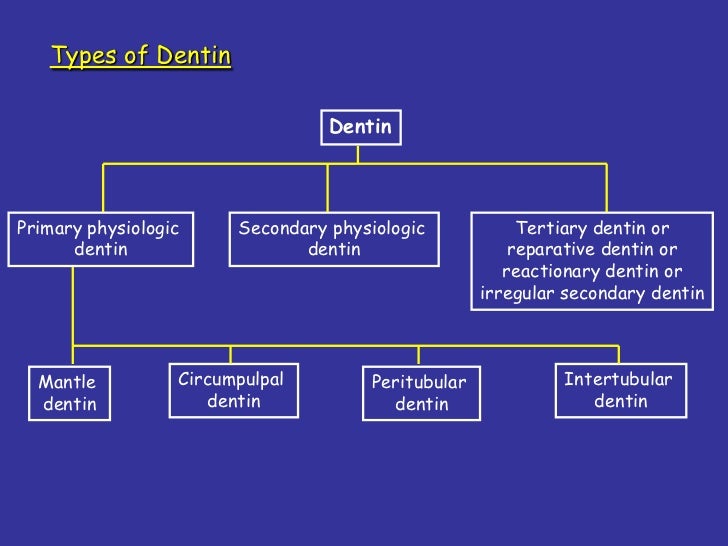
Types of Dentin
Hey friends,
Dentin is a very important question.
It comes as a question worth 4 points in my theory exam paper! I have tried my best to make it simpler for you all in this blog :))
I hope this will help you!
A. Mantle Dentin
Abrasion
Erosion
Cavity preparation
Written by Anisha Valli :))))
Primary Dentin
It is divided into Mantle and Circumpulpal Dentin
A. Mantle Dentin
- First formed dentin in the Crown
- Type III collagen
- It is less mineralized
- Matrix vesicles are present which help in Globular calcification
- It forms the bulk of the tooth
- Type one collagen
- It is more mineralized
- Matrix vesicles are present which help in Linear and globular calcification
Secondary Dentin
- It is formed after the root completion
- It contains dentinal tubules which are S-shaped
- The mineral ratio is similar to primary Dentin
- Secondary Dentin is a narrow band of Dentin bordering the pulp
- As age increases, inorganic content increases
- Therefore the Dentin becomes sclerosed
- It means It protects the pulp from exposure in older teeth
Tertiary Dentin
It is formed in response to stimuli
AttritionAbrasion
Erosion
Cavity preparation
- It is deposited on the pulpal surface of Dentin only in the affected area
- The appearance of Dentin varies as it is formed by an odontoblast
- Quality and quantity of tertiary Dentin depends on intensity and duration of stimuli
Reactionary Dentin
Dentin is deposited by pre-existing odontoblasts
Reparative Dentin
Dentin is deposited by newly differentiated odontoblast-like cellsWritten by Anisha Valli :))))
Sunday, February 4, 2018
Transamination
Have you ever wondered about the difference between non-essential and essential amino acids?
I’m pretty sure you know the difference :))
If non-essential amino acids are not delivered to the body through diet then how are they made in the body?
Answer is simple it is by the process of transamination
I hope my notes will help you! If you have any doubts, don’t hesitate to comment or send a message on WhatsApp group :)


Maxillary Artery notes
Hello Friends! This is Anisha :))
Maxillary artery is divided into three branches. Again, each branch is subdivided.
We also have to learn their course which is very confusing and we forget it during our exam :(
So, I came up with an easy way to learn it. I decided that I will show the course of the artery in form of a diagram ( you will get more marks! ) and write down what it supplies.
I hope my notes will help you :)) All the best
Subscribe to:
Posts (Atom)