Showing posts with label Dermatology. Show all posts

Friday, April 21, 2017

Nail Changes in Medicine : A Summary

Hi everyone. Just a list of changes you can see in the nails in different systemic Diseases. So let's get nailed ;)

1. Clubbing -
Loss of angle between the nail and the nail fold - More soft and bulbous nail.
Typically indicates Cardio Pulmonary function disturbance :
--> Cardiac conditions like Cyanotic heart disease, Infective endocarditis and Atrial myxoma.
--> Respiratory conditions :
Neoplastic like CA lung ( Esp. Squamous cell CA) , Mesothelioma.
Infective like Bronchiectasis , Abscess , Empyema.
(Non cardiorespiratory causes = Inflammatory bowel disease, Biliary Cirrhois.
Thyroid Acropachy , Acromegaly. )

2. Koilonychia -
Spoon shaped nails.
Strongly indicative of Iron Deficiency anemia or Fungal nail infection.

3. Onycholysis -
Destruction of nail. 
Seen in Psoriasis , Hyperthyroid and Fungal nail infection.

4. Chronic Paronychia -
Inflammation of nail fold. May have swollen nail and discharge with throbbing pain. May occur due to frequent nail biting.

5. Cyanosis -
Can be looked for in nail bed. We have a post on this already.

6. Beau line -
Transverse furrows from temporary arrest of nail growth due to increased stress.
Nails grow at 0.1 mm/d , so furrow distance from the cuticle can be used to time the attack. Can be seen in Malaria , Typhus , Rheumatic fever , Kawasaki.

7. Mees line -
White transverse bands in Arsenic poisoning / Renal failure.

8. Muerhcke's line
White parallel lines without furrowing on the nail.
Seen in Hypoalbuminemia.

9. Terry's nails -
Proximal portion of nail is white / pink , tip is reddish brown.
Seen in cirrhosis , CRF

10. Splinter hemorrhage -
Longitudinal Hemorrhage streaks under the nail seen in Infective endocarditis.

What a fun way to get nailed down 😂 Happy studying !
Stay awesome.

~ A.P.Burkholderia.

Thursday, April 20, 2017

Edge of an ulcer : An overview

Hello

An overview on how edge of an ulcer appears with characteristic identification features depending on the underlying causes: (SPURE)

Sloping edge - Venous ulcer, also seen in traumatic cases. It is red - purplish in color and consists of new healing epithelium. ( spreading type )

Punched out edge - Arterial and Neuropathic ulcer. Edges are punched out at right angles. ( non spreading type )

Undermined edge - Decubitus and Tuberculous ulcer. It spreads rapidly to destroy the surrounding tissue !!

Rolling back - Basal cell Ca. It is characterised by raised, pearly white beaded edge with central necrotic tissue.

Everted edge - Squamous cell Ca. It is a rapidly growing invasive ulcer with heaped up and everted edges.


Thats all
Hope this helps :)

- Jaskunwar Singh

Saturday, January 14, 2017

Artificial skin

Hello awesomites !!! Today's topic is something that really has created revolution in the entire human race - "Artificial skin"

Skin is the largest organ of our body. It consists of three parts Epidermis, dermis and fatty layer. Epidermis serves as the protective layer of the skin, it prevents the entry of the pathogen into body with the help of sebaceous and sweat glands along with long chain fatty acids, and dermis consists of all the nerves, artery and veins that supply skin. Fatty layer maintain the body temperature.
A damage to the skin through burns causes loss of large number of plasma and it provides free entry to pathogens into body. Artificial skin was discovered by Loannis yannas & his colleague surgeon Dr John burke.

Artificial skin was discovered to replace bandages, which cannot seal large damage areas. Patients with extensive burns often die as bandages cannot heal their wounds completely. Even if patient survives some scars are left behind on skin. Artificial skin consists of collagen polymers. Collagen is protein found in skin. Artificial skin helps patient to grow skin without forming any scars. It has been commercially prepared under name as IntegraTM.

It is also used in plastic surgery and also in chronic skin wounds.
 
Ahh!    That's it :)
            Keep smiling :)
            Have a nice day!
      
~Ojas

Wednesday, December 21, 2016

Bullous skin disorders-1 pemphigus vulgaris and bullous pemphigoid

Pemphigus vulgaris


http://www.medpictures.org/pemphigus-vulgaris-pictures.html

Bullous pemphigoid


http://www.pcds.org.uk/clinical-guidance/bullous-pemphigoid1#!prettyPhoto


these two are the most commonly asked questions within bullous diseases.
the major difference you need to know is
pemphigus vulgaris- bullae are suprabasal. hence these are not tense and rupture easily..not very common in a clinical case for you to find a bulla
bullous pemphigoid- bullae are subepidermal..and hence tense.

a mnemonic..like a bull..bullae in Bullous pemphigoid are tough and stronger

-sakkan

Wednesday, December 7, 2016

Stigmata of infective endocarditis

Hey Awesome people!

Here is another stigmata apart from those mentioned previously by IkaN in a post here,

Stigmata of infective endocarditis:-
(Remember- It's all in the hands)

Janeway's lesions: non- tender, erythematous macules on the palms and soles.
Osler's nodes: ephemeral spots of painful nodular erythema on hands and feet.
- Splinter haemorrhages: seen on the nails.

Thats all!

- Jaskunwar Singh

Tuesday, August 16, 2016

Different types of skin lesions (as in rashes)

                          Hey guys, we started Internal Medicine Last week, so we were studying about different types of skin lesion as in rashes in Harrison’ Internal Medicine 19e. So I made little chart myself to memorize it. So I thought to share it with you all.

Monday, July 11, 2016

Treatment of acne mnemonic

Hello!

Here's a short post on acne.

Antimicrobials for treatment of acne mnemonic: ABCDE
Azelaic acid
Benzoyl peroxide
Clindamycin
Dapsone
Erythromycin

Monday, May 2, 2016

Sturge- Weber syndrome

Hello

Talking about this rare syndrome, I read about in ophthalmology lecture (class of secondary glaucomas) today... So let's start with it-
- It is also known as Encephalo Trigeminal  Angiomatosis (ETA)
• Rare congenital Neurological and skin disorder (phakomatoses)
• Caused by Somatic Acivating Mutation in GNAQ gene.

Port -wine stains (nervus flammeus) - 
            ○ Usually seen on Forehead and Upper Eyelid of one side of face; present since birth.
            ○ Light Pink to Deep Purple
            ○ Caused by Overabundance of capillaries  around Ophthalmic branch of Trigeminal nerve.

• Associated with
          ○ Secondary Glaucoma (in 50% patients)
          Buphthalmos (enlarged eyeball due to increased intraocular tension)
          ○ Leukocoria (white pupillary reflex)
          Neurologic manifestations- seizures, convulsions (on side of body opposite to birth mark), mental retardation, calcification of tissue and loss of nerve cells in cerebral cortex.
          Ipsilateral Leptomeningeal Angioma (on same side of birth mark; calcification of underlying brain and atrophy of affected region)
Malformed blood vessels in the piamater overlying the brain on same side of head as the birthmark.

Radiological appearance shows Tram-Track Appearance on CT, bilaterally.

Treatment strategies include Laser surgery, Hemispherectomy
 - Latanoprost, a Prostaglandin analogue, is suitable drug for decreasing intra ocular pressure. (1 drop daily in evening)

That's all
- Jaskunwar Singh

Tuesday, December 4, 2012

Dermatophytes

What are Dermatophytes?
They are a group of filamentous fungi that infect only the superficial keratinized tissues
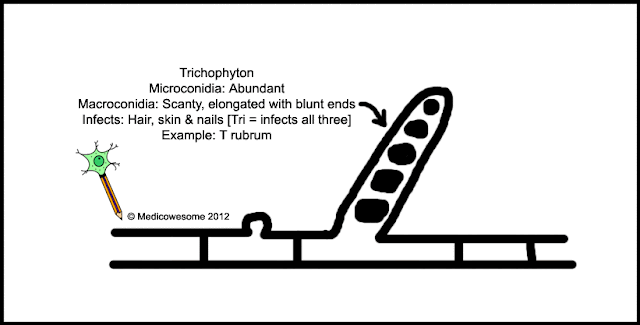
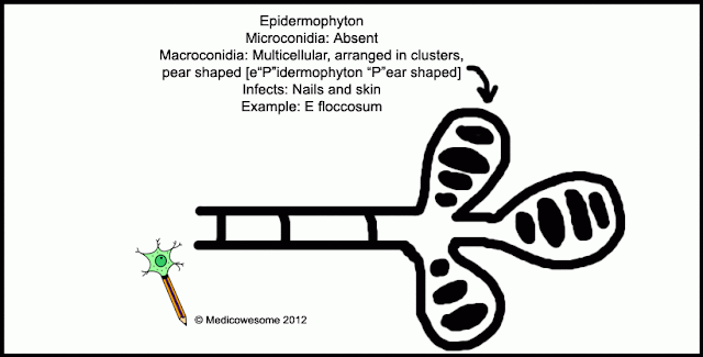
They are classified into three genera: Trichophyton, Microsporum & Epidermophyton
I had a tough time learning them at first so I made a mnemonic ^_^"
I have put it up in the image itself

Trichophyton mnemonic
Microsporum mnemonic

Epidermophyton mnemonic


Did you know?
Fungal Wet Mount is used for direct detection of fungal forms in patient specimens
KOH lyses host cells and keratin and makes fungal elements more easily detected by elimination of host materials
Calcofluor white, a fluorogenic dye, binds to specific polysaccharide bonds found in the chitin-rich fungal cell walls and can be used for detection of fungi too ^_^

That's all =D

-IkaN